| MULTI-MODALITY RESEARCH: ONDAMED® Biofeedback/PEMF & the Doppler Ultrasound August 5, 2022 Many visionaries in health and medical innovations collaborate on the expansion of their work through continued exploration and referencing from other technologies. Two such innovators have shared countless hours exchanging notes early this year, to finally meet at this special crossroads of scientific exploration. Dr. Silvia Binder (founder of the Binder Institute for Personalized Medicine in So. Germany) and Dr. Robert Bard (seasoned ultrasound researcher and biometric imaging validator) conducted an exploratory performance review through the integration between their respective non-invasive technologies. The innovation in review is the ONDAMED®- a full-body biofeedback device + PEMF therapeutic solution designed to target the root causes of physiological imbalances such as pain, injuries, inflammation and neurological disorders. According to Dr. Binder, this energy therapeutic device has a major global following, recognized in areas including sports medicine, pain management, psychiatry, anti-aging and neurology. This validation project which started in July 8 was designed to confirm the vast number of testimonials about its advantages including (but not limited to) pain & stress reduction, optimizing mental and emotional wellness and balancing hormones and metabolism. This research also hopes to challenge or confirm its ability to support detoxification, enhance cellular nutrient absorption, inflammation reduction, fighting off infections, cellular repair/regeneration, improve the immune function and hemodynamics / circulation. This exploratory validation project is co-directed by Dr. Bard from his NYC research facility through the use of his various advanced ultrasound models. Combining the diagnostic prowess of the ultrasound has become Dr. Bard's preferred choice for imaging because of its (safe) non-radiation and real-time properties- and added to this is its ability to perform near and within other electronic regenerative technologies (like the ONDAMED® Biofeedback and PEMF) without any interference. This three-day scientific review is a testament of strategic vision between health innovators aspiring to forge and confirm new answers while fostering the non-invasive medical technology movement. Where both the ONDAMED® device and the Ultrasound are two of the latest in non-surgical medical marvels, combining the unique diagnostic abilities of biofeedback and the imaging validation of ultrasound (through quantitative biometrics) reading the effects of the PEMF therapeutic functions of the ONDAMED® offers a new way to support, analyze and record evidence of regenerative medicine. (Below is Part 1 of this integrative research project) |
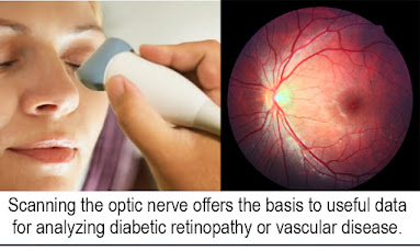
SCANNING THE OPTIC NERVE
Written by: Dr. Robert L. Bard
This study is part of an ongoing review of the quantifiable scanning features of the high frequency ultrasound probe w/ 3D Doppler- assessing the biometrics of a biofeedback electromagnetic device. This review aims to form the basis for analyzing cases such as diabetic retinopathy or hypertensive vascular disease. Secondary protocols for treating disease due to Alzheimer's or chronic trauma by increasing the blood flow in the brain to prevent further damage may be possible based on the imaging findings derived from this research.
The subject complaining of headaches had a prior case of malaria (which is a small parasite that lodges itself throughout the body) demonstrated benign lymph nodes in multiple areas in the head and neck. We opted to start by scanning ocular orbit to detect if increased intracranial pressure on the optic nerve head was detectable to verify potential cerebral pathology
Initial scans show the largest diameter was 0.5mm and we also found a benign microcalcific deposit. We are searching for sensitive blood vessels to look for obvious changes in the retina. Next, we introduced the biofeedback energy applicator closer towards the eye. Here, we notice (through our ultrasound readings) that the vessels have dilated about 30% during the treatment.
The measurement of the vessels post-treatment with blood flow and with the eye in the same position, showed that the overall vascularity had increased or became more visible to the blood flow technology. This means that the resting vessels had expanded from 0.5mm to 0.7 and as far up to 0 .9mm. This translates to roughly a 30-40% increase in the blood flow, going to the area that was exposed to electromagnetic stimulation.
Notice the dark band at the bottom center which represents the optic nerve. Two things we note: (1) there's no bulging of the optic nerve disc, which means that the intracranial pressure is normal. (2) Also at the tip of the optic nerve, there is a white vertical object, like a small thumbprint -and to the left of that is some vascular flow. This is a sub millimeter calcific area at the optic nerve head, which is called a DRUSEN. It's a benign finding that may be linked to increased atherosclerosis.
In comparison to the BEFORE clip, prior to being exposed to biofeedback and energy treatment transmission that the small blue and orange vessels are much larger this time, indicating observable realtime effect of treatment.
The buildup of toxins in the cells can decrease your health and wellness. Cell phones, computers, TV, air quality, improper hydration, and poor-quality foods can all lead to poor cellular health. When treatments that help detoxify, the body initially occurs occasionally this is when the Herx Reaction occurs. Herx reaction is not common but can occur. Education and communicating to the user on the possibility will improve outcomes and the persons reaction. These symptoms peaks eight hours after treatment and disappears normally within 24-36 hours.
SEE COMPLETE REVIEW ON HERXHEIMER'S REACTION
- As part of Heart Health Awareness Month, the Integrative Pain Healers Alliance , the Angiofoundation , the editors of Prevention101 and N...
- By: Jessica Connell-Glynn, LCSW Edited by: Roberta Kline, MD Health issues are often linked to a range of emotional distress, but a major c...
- IPHA NEWS and Health & Healing 101 takes you across our northern border to Quebec, Canada for an up close interview with a leading thera...
Disclaimer: The information (including, but not limited to text, graphics, images and other material) contained in this article is for informational purposes only. No material on this site is intended to be a substitute for professional medical advice or scientific claims. Furthermore, any/all contributors (both medical and non-medical) featured in this article are presenting only ANECDOTAL findings pertaining to the effects and performance of the products/technologies being reviewed - and are not offering clinical data or medical recommendations in any way. Always seek the advice of your physician or other qualified health care provider with any questions you may have regarding a medical condition or treatment and before undertaking a new health care regimen, never disregard professional medical advice or delay in seeking it because of something you read on this page, article, blog or website.


.JPG)


