Friday, June 24, 2022

DERM-NEWS 2022: TOXINS IN THE SKIN

According to the CDC, DERMAL ABSORPTION happens when a chemical goes through the skin and travels into the body. Many chemicals used in the workplace or even from home (ie. pesticides and organic solvents) can wreak havoc on and under the skin, damage internal organs and also the immune system if they penetrate the skin and enter the bloodstream. Most efforts to address chemical hazards have been focused on breathing, digesting or drinking chemicals rather than what's being absorbed through skin. Because of this, there are far fewer methods and campaigns dedicated to assessing skin exposures to toxins - UNTIL NOW.

With the latest evidence of inflammatory skin disease and foreign bodies under the skin, the medical and aesthetics communities are now facing the many new ways that environmental (toxic) influencers can affect the body through skin contact and absorption. This initiative is achieved thanks to our non-invasive diagnostic imaging advancements including Confocal Microscopy and the 3D Doppler Ultrasound. Dr. Robert Bard, presents this critical health topic as part of his 2022 Series on INTRADERMAL IMAGING OF TOXINS in dermatology conferences such as the NIDISKIN Spring Symposium. (See INFLAMMATORY SKIN DISEASE Poster)

SUPPLEMENTAL:
 Toxins and Health: A Personalized Medicine Perspective: By Dr. Roberta Kline


SENSIBLE ADVANTAGES OF ULTRASOUND IMAGING FOR BURNS AND DERMAL TRAUMAS

Written by: Dr. Robert L. Bard of the Bard Diagnostic Imaging Center

For most emergency responders and physicians, identifying the degree of any burn or dermal trauma cases starts with a visual assessment.  With professional training and enough experience, the professional eye can differentiate between first, second and third degree burns to initiate the proper treatment process. First-degree burns commonly show redness, and swelling only on the outermost skin layer; second-degree burns show surface injury to the underlying layer with blistering and Third-degree burns affect up to the deep layers of the skin.


As standard practices continue to evolve, diagnosing any advanced burns are now calling for new considerations for the prevention of burn-related complications.  As more and more after-effects from high degree burns have left patients with lasting (and sometimes fatal) results, it may no longer be enough to drive a treatment protocol based on surface topical healing. 

BURN SCANNING TECHNOLOGY: ASSURANCE AGAINST COMPLICATIONS
Second and third-degree burns may show blisters and red skin but with today’s many non-invasive subdermal technologies, you can now identify the depth of the burn and what the injury truly means under the skin. Identifying the exact DEPTH of the internal injury as well as monitoring (visually) its internal impact/effect on the body may uncover and predict other potential health issues such as:

-        Scarring and unrecoverable dead tissues
-        Damage to Nerve endings /neuropathy
-        Inflammation
-        Temporary to permanent loss of skin
-        Damage to underlying bones, muscles and tendons
-        Bacterial infections (from the broken skin) like tetanus
-        Internal Shock
-        Hypovolemia (low blood volume/ unusual blood loss from a burn)

According to Image 1A, high resolution sonogram used a standard probe for skin imaging showing the black area, which is just below the white line of the surface and the black fluid corresponds to the blister seen on the specimen of the burned area. Now below it you see the skin with the first vertical blue dotted line and it goes to the fascial plain white line, which shows swelling of the tissues. The normal skin measures 1.3 millimeters or thin as a dime and the depth of the burn itself measured is twice that, 2.6 millimeters. So we have a way of seeing the fluid. We have a way of checking the depth of the burn, which is clinically difficult because the eyes cannot see below the skin.   You can see the blister is basically gone because that's the tiny black area above the tissue line- showing almost no fluid left (represented In the blackish area)


Upon review of Day 9 diagram, see "burn healing" on the left side of this diagram where it says dermis on day 9, you can see the bottom white line under the lettering dermis, which shows the bottom of the skin, which last time was 1.3 millimeters. Then to the right of that the dermis tissue is starting to have the red and blue healing blood vessels that's coming in marked by the red arrow. And then to the right of that where it says decreased vascularity, the larger blood vessels have not yet come in, but at least we know the skin has viable feeding blood flow. So it's more likely than not to heal.

** These images are scanned with the GE Ultrasound Voluson E8.  Any machine over 15 megahertz can be used on burns- however, devices with higher the resolution improves the scan experience to get the best data.

REVIEW OF THE BLOOD FLOW INNOVATION
Today’s imaging devices cover a wide range of functions on the market carrying specific features to fit their many users specific needs.  This scan was generated by the General Electric Voluson E8 system which uses an 18 mhz probe outputting 1/10 of a millimeter of resolution.  The benefit of using this GE 3D Doppler system enables the ability to measure the depth of the burn as well as identify and record the exact amount of fluid in the surface of the burn, which is the blister. 

For many diagnostic applications, the real-time scanning ability of VASCULAR ULTRASOUND has greatly advanced the way injuries are read, identified and managed.  Vascular ultrasound uses sound waves to evaluate the body's circulatory system.  It also helps identify blockages in the arteries and veins and detect blood clots.  This innovation is not radiation based, leaving no harmful side effects and out-performs many of today’s current counterparts including accuracy in scanning soft tissues that does not appear in x-rays. 
BLOOD FLOW technology is the “diagnostician’s storyteller”.  It allows you to see which part of the skin is alive by the indication of active blood flow through the area- versus the skin that is dead or dying with no blood flow. In cases of burns, the margin of injury can extend once the burn has been completely cooled down. Since vascular ultrasound is safe sound waves, you can conduct frequent scans to monitor healing and progress every hour/every day until resolution.

The paradigm of studying blood flow allows any diagnostician to review whether the tissue is curing or not.  In the case of performing a possible skin graft, the blood flow around an injury gives more data as far as the behavior of the burn or injury as well as the condition of healthy tissue to attach to the burn.

Any inflammatory skin disease is caused by inflammatory blood vessels, which is not evident by the naked eye. This scanning modality allows you to quantify the degree of inflammation and the response to all the new treatments available. Where widely accepted optical technologies work well,  they are limited to 1/2 of a millimeter depth, so they are surface only.


SKIN LIFE VS. SCARS 
If the tissue doesn't heal with normal skin, it will scar. The scar tissue appears as black in ultrasound imaging, almost like the fluid- but with zero blood flow.  Any kind of trauma can result in healing tissue or dead tissue, which will either get infected or scar down. Imaging can also show if the area is getting inflamed as it indicates irregular volume of the blood vessels resulting in cellulitis or the inflamed skin.

Scar tissue is dead skin.  Doppler Imaging can be useful as it shows the thickness of the scar to determine if it can be treated, either with steroids, laser or any of a number of current scar treatment technologies.  The depth and the hardness of the scar determines which option to use and all these can be resolved by the various ultrasound technologies. Ultrasound is the new ‘weapon of choice’ to show depth, thickness of the scar, type of scar, how hard or elastic it is  (also see elastography).  It also allows the surgeon to clearly identify the margins you wish to attach the graph to.  


[IMAGE 2] In this image, we have a burn that came from a charcoal grill. This burn leaves a white coating (surface singe) to the red skin. (A) This white surface outline with the black arrows is the ash from the grill or the burning surface. The small yellow circle is the blister that immediately broke from the heat. So the blister burst and opened up causing the teardrop-shaped opening in the skin, which could get infected. 
Diagram B shows the two yellow arrows pointing to the white area, it's got a top white, a medium dark, and a bottom white area.  That's the appearing ash visible only on the surface but not penetrating deep (thus it is not a third-degree burn). Upon further interactive review of the burn, it was only surface ash from the surface of the charcoal grill which was easily removable. On the same image (B), we are also looking at external tendon, 1 mm wide. 
Diagram C indicates the blood vessels and the normal tissue on the side of the burn. Though the burn goes deep into the skin, it is not a complete third-degree burn in the whole area (B). Comparing B and C, the injury to the burned tissue is marked by the red arrow on top and also the tendon that raises the finger pushes 1 mm wide is completely unaffected by the dark burn area. Now below that since we weren't sure if it was a third-degree burn or we wanted to see if there was viable skin next to it, we did the blood flow technology which shows the micro vessels or the capillaries that are in the adjacent skin, so if you ever needed to graft it you'd have normal skin and also the fact that you have normal skin in the red area means that the burn in that area is a first degree or not really burned at all.

“IT’S ALL ABOUT THE PROBES”
The GE Doppler scanner can go deeper under the skin - at an estimated 5x the resolution than the average ultrasound probe (at 1/10mm resolution). The higher the megahertz, the deeper and sharper the image (like 70mhz has 1/50 of a millimeter resolution).  Such a probe is much better for imaging tendons and skin and the regular 18 or 20 megahertz (such as the GE) that we use routinely use has 1/10 of a millimeter resolution. You have better detail for seeing tendons and blood vessels.

Overall, each probe determines a specific depth, the width & range of the scan, the level of blood flow while the hardware & diagnostic software itself communicates with the probe to translate all data into recognizable images in real time.

PRE-OPERATIVE (AND RISK REDUCTION) PROTOCOL
Among its many uses, cosmetic surgeons can benefit from dermal imaging by mapping the nerves and the arteries before cutting. Also, you can find the dead skin as compared to the normal skin for doing reconstructive surgery. 

Emergency departments can more easily treat nerve trauma, burns, tendon injuries with the help of visual analysis of any affected area.  As an example,  you can see if the tendon is partly or completely torn with ultrasound more easily and effectively. you are able to move the finger because it won't be any movement of the torn part. If you move the tendon when you open up the finger from a closed fist position.


ABOUT THE AUTHOR-

ROBERT L. BARD, MD, PC, DABR, FASLMS - Advanced Imaging & Diagnostic Specialist
Having paved the way for the study of various cancers both clinically and academically, Dr. Robert Bard co-founded the 9/11 CancerScan program to bring additional diagnostic support to all first responders from Ground Zero. His main practice in midtown, NYC (Bard Diagnostic Imaging- www.CancerScan.com) uses the latest in digital Imaging technology has been also used to help guide biopsies and in many cases, even replicate much of the same reports of a clinical invasive biopsy. Imaging solutions such as high-powered Sonograms, Spectral Doppler, sonofluoroscopy, 3D/4D Image Reconstruction and the Spectral Doppler are safe, noninvasive, and does not use ionizing radiation. It is used as a complement to find anomalies and help diagnose the causes of pain, swelling and infection in the body’s internal organs while allowing the diagnostician the ability to zoom and ‘travel’ deep into the body for maximum exploration.


Disclaimer & Copyright Notice: The materials provided on this website are copyrighted and the intellectual property of the publishers/producers (The NY Cancer Resource Alliance/IntermediaWorx inc. and Bard Diagnostic Research & Educational Programs). It is provided publicly strictly for informational purposes within non-commercial use and not for purposes of resale, distribution, public display or performance. Unless otherwise indicated on this web based page, sharing, re-posting, re-publishing of this work is strictly prohibited without due permission from the publishers.  Also, certain content may be licensed from third-parties. The licenses for some of this Content may contain additional terms. When such Content licenses contain additional terms, we will make these terms available to you on those pages (which his incorporated herein by reference).The publishers/producers of this site and its contents such as videos, graphics, text, and other materials published are not intended to be a substitute for professional medical advice, diagnosis, or treatment. For any questions you may have regarding a medical condition, please always seek the advice of your physician or a qualified health provider. Do not postpone or disregard any professional medical advice over something you may have seen or read on this website. If you think you may have a medical emergency, call your doctor or 9-1-1 immediately.  This website does not support, endorse or recommend any specific products, tests, physicians, procedures, treatment opinions or other information that may be mentioned on this site. Referencing any content or information seen or published in this website or shared by other visitors of this website is solely at your own risk. The publishers/producers of this Internet web site reserves the right, at its sole discretion, to modify, disable access to, or discontinue, temporarily or permanently, all or any part of this Internet web site or any information contained thereon without liability or notice to you.


Wednesday, June 22, 2022

HERXHEIMER'S REACTION: REVIEWING THE HEALING CRISIS

"HERX IS NOT A SIDE EFFECT!"

By: Josh Schueller, PT


As non-invasive treatments such as electromagnetic and neuro-stimulation devices have shown significant evidence as a safe and effective option for wellness and pain relief within recent decades, one possible bodily reaction may occur that can feel adversely as pain or discomfort- possibly being misunderstood as a side effect of the treatment.  This physiological reaction is called Herxheimer Reaction. This can occur when dead microbes or bacteria release endotoxins into your body at an accelerating rate.   This accelerated release is quicker than the body can eliminate.  The body then initiates an immune response which can bring on an inflammatory response.  Otherwise known as "Herk reactions", this reaction is commonly a temporary and short term (normally only last a couple hours to days) detoxification reaction in your body.  Common symptoms can resemble the flu- headache, joint and muscle ache, malaise, chills, nausea, sore throat etc.   This reaction is caused by the detoxification of the body.  Staying hydrated is one way to help flush your body and decrease the symptoms.   

A TEMPORARY REACTION TO DETOXING
The buildup of toxins in the cells can decrease your health and wellness.  Cell phones, computers, TV, air quality, improper hydration, and poor-quality foods can all lead to poor cellular health.  When treatments that help detoxify, the body initially occurs occasionally this is when the Herx Reaction occurs. Herx reaction is not common but can occur.  Education and communicating to the user on the possibility will improve outcomes and the persons reaction.  These symptoms peaks eight hours after treatment and disappears normally within 24-36 hours. 

As with any new exercise keeping your cells hydrated and healthy is the best remedy.  Other helpful ways to lessen the effects is to get plenty of sunshine, minimize exercise during this time, keeping your diet pure (avoiding alcohol) and using a sauna or sea salt bath. Herx Reactions more commonly occur if during the first few treatments the intensity and/or duration is too high for the treatment.  As with any new activity, starting low and slow and incrementally increasing intensity and duration is essential to avoiding any unwanted effects.  Some indications for a possible Herz reaction are how long the person has suffered from the condition.  Chronic conditions can demonstrate more likely increase in the amount of toxins in the system and poor cell health.  If a Herx reaction occurs- decreasing the intensity and duration for a couple of treatments is the most appropriate action needed.  


PART 2: THE HEALING CRISIS
By: Dr. Roberta Kline


Introduction
In health care, it is common for physicians to witness manifestations of what is commonly referred to as a “healing crisis”.  This experience can appear in many different ways, including physical, emotional and spiritual symptoms that are then followed by a greater sense of well-being. While many “healing crises” have been in response to treatment of Lyme disease and Candida, nutritional changes also occur - from “detox” programs and energetic healing.  There may be lower rates of healing crises within less aggressive and more personalized interventions, but the condition seems to exist here too. 

This phenomenon is not widely recognized within the conventional medical community. Even among complementary and alternative medicine (CAM) practitioners, where there is more recognition of this phenomenon, it is still poorly understood.  Through the course of scientific evolution, we hope to find an answer to whether it is possible to better predict predisposition to experiencing a healing crisis or Jarisch-Herxheimer Reaction.  If so, perhaps we can develop personalized treatments to minimize these symptoms while still creating an effective healing response.


THE JARISCH-HERXHEIMER REACTION

First described in relation to treatment of a syphilis patient by the Austrian dermatologist Adolf Jarisch in the late 1800’s (and similarly reported by German dermatologist Karl Herxheimer), the Jarisch-Herxheimer Reaction (JHR)  has been reported with treatment of other spirochete infections, including leptospirosis, Lyme disease, and relapsing fever, as well as with some fungal, protozoan and bacterial infections. Abridged as the Herxheimer's (or Herx) Reaction, it is today's best-known example of a healing crisis.  This transient phenomenon occurs within 24 hours of antibiotic or antifungal treatment, and often includes fever, chills, headache, nausea and vomiting, tachycardia, hypotension, myalgia and exacerbation of skin lesions. It is not a rare reaction, occurring in over 50% of patients in some studies. While supportive care is indicated, it is rarely life-threatening. 

While efforts to understand the underlying mechanisms have focused on scientific studies related to spirochete infections, the exact pathophysiology still remains elusive. Theories have mostly focused on the concept of “die-off” – that a sudden burden of dead spirochetes releases toxins and induces an inflammatory reaction. (1)

More recent research has expanded to include B. Burgdorferi, the spirochete associated with Lyme disease, and better technology has shed some additional light on the process underlying this phenomenon. No longer is the theory of spirochete “die-off” considered a primary trigger, as the symptoms appear long before the antibiotics actually result in death of the organisms.

It appears to be mediated by a multifactorial inflammatory process, potentially provoked by the uptake of the spirochete into the polymorphic neutrophils. This complex inflammatory response likely also includes nonendotoxin pyrogen and organism-specific lipoproteins, that then trigger an inflammatory cascade driven by tumor necrosis factor alpha (TNF-alpha), and downstream cytokines including interleukin 6 (IL-6) and interleukin 8 (IL-8). But it is still not clear how much of this inflammatory response is causal, and how much is the result of the Jarisch-Herxheimer reaction, and further research is needed. (2)

Interestingly, reports of a condition known as cytokine release syndrome are emerging as a result of treatment with CAR T-cell therapy, which is a form of immunotherapy for cancer. The symptoms occur within hours to days, and are very similar and can include fever, tachycardia, hypotension, and rash. The syndrome is thought to be caused by a large and rapid influx of cytokines – very similar to the findings from studies on the JHR. (3)


CROSSROADS BETWEEN PAIN AND WELLNESS

Various other healing modalities have also reported responses to treatment called “healing crises” that appear to be similar to JHR. Although many people refer to them generically as Herxheimer reactions (“herxing” for short), they do have some differences. Often pain, including reactivation of “old” pain, is seen in addition to changes in heart rate, respiration, more frequent urination and bowel movements, fatigue, fever, headaches, skin rashes and/or disrupted sleep. It is believed that many of these symptoms are the body’s way of eliminating accumulated waste products and toxins that are now being released as a result of the therapy. (4)

Even the reactivation of pain in locations that had not bothered the person for years is seen as part of this process. Oftentimes the healing crisis is followed by improvement in emotional and spiritual wellbeing, in addition to physical health. But while this phenomenon is recognized by many CAM practitioners, there is very limited research available and most of our understanding is anecdotal. Much more is needed to enhance our knowledge so we can educate both healthcare professionals as well as their patients. 


References:

[1] Aayush Dhakal and Evelyn Sbar. Jarisch Herxheimer Reaction. StatPearls April 28, 2022.

[2] Thomas Butler. The Jarisch–Herxheimer Reaction After Antibiotic Treatment of Spirochetal Infections: A Review of Recent Cases and Our Understanding of Pathogenesis Am J Trop Med Hyg 2017 Jan 11; 96(1): 46-52

[3] Noelle Frey and David Porter. Cytokine Release Syndrome with Chimeric Antigen Receptor T Cell Therapy. Biol Blood Marrow Transplant. 2019 Apr;25(4):e123-e127

[4]  Thora Jenny Gunnarsdottir and Helga Jonsdottir. Healing crisis in reflexology: Becoming worse before becoming better. Complementary Therapies in Clinical Practice 16 (2010);239-243


Tuesday, June 14, 2022

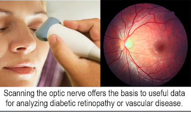
BIOFEEDBACK & ELECTROMAGNETIC EXPOSURE: SCANNING THE OPTIC NERVE


MULTI-MODALITY RESEARCH: 
ONDAMED
® Biofeedback/PEMF & the Doppler Ultrasound
August 5, 2022

Many visionaries in health and medical innovations collaborate on the expansion of their work through continued exploration and referencing from other technologies.  Two such innovators have shared countless hours exchanging notes early this year, to finally meet at this special crossroads of scientific exploration.  Dr. Silvia Binder (founder of the Binder Institute for Personalized Medicine in So. Germany) and Dr. Robert Bard (seasoned ultrasound researcher and biometric imaging validator) conducted an exploratory performance review through the integration between their respective non-invasive technologies.  

The innovation in review is the ONDAMED®- a full-body biofeedback device + PEMF therapeutic solution designed to target the root causes of physiological imbalances such as pain, injuries, inflammation and neurological disorders. According to Dr. Binder, this energy therapeutic device has a major global following, recognized in areas including sports medicine, pain management, psychiatry,  anti-aging and neurology.  This validation project which started in July 8 was designed to confirm the vast number of testimonials about its advantages including (but not limited to) pain & stress reduction, optimizing mental and emotional wellness and balancing hormones and metabolism.  This research also hopes to challenge or confirm its ability to support detoxification, enhance cellular nutrient absorption, inflammation reduction, fighting off infections, cellular repair/regeneration, improve the immune function and hemodynamics / circulation.

This exploratory validation project is co-directed by Dr. Bard from his NYC research facility through the use of his various advanced ultrasound models.  Combining the diagnostic prowess of the ultrasound has become Dr. Bard's preferred choice for imaging because of its (safe) non-radiation and real-time properties- and added to this is its ability to perform near and within other electronic regenerative technologies (like the ONDAMED® Biofeedback and PEMF) without any interference. 


This three-day scientific review is a testament of strategic vision between health innovators aspiring to forge and confirm new answers while fostering the non-invasive medical technology movement. Where both the 
ONDAMED® device and the Ultrasound are two of the latest in non-surgical medical marvels, combining the unique diagnostic abilities of biofeedback and the imaging validation of ultrasound (through quantitative biometrics) reading the effects of the PEMF therapeutic functions of the ONDAMED® offers a new way to support, analyze and record evidence of regenerative medicine.  
(Below is Part 1 of this integrative research project)


SCANNING THE OPTIC NERVE

Written by: Dr. Robert L. Bard

This study is part of an ongoing review of the quantifiable scanning features of the high frequency ultrasound probe w/ 3D Doppler- assessing the biometrics of a biofeedback electromagnetic device. This review aims to form the basis for analyzing cases such as diabetic retinopathy or hypertensive vascular disease.  Secondary protocols for treating disease due to Alzheimer's or chronic trauma by increasing the blood flow in the brain to prevent further damage may be possible based on the imaging findings derived from this research. 

The subject complaining of headaches had a prior case of malaria (which is a small parasite that lodges itself throughout the body) demonstrated benign lymph nodes in multiple areas in the head and neck. We opted to start by scanning ocular orbit to detect if increased intracranial pressure on the optic nerve head was detectable to verify potential cerebral pathology

 INVESTIGATING ELECTROMAGNETIC TECHNOLOGIES
Our study team has been following the effect of electromagnetic devices including a Biofeedback and targeted electromagnetic stimulator (Ondamed®) since this non-invasive treatment device is promoted to support pain relief, soft tissue injuries and wound healing and reportedly shows success in providing clinical effects such as: anti-inflammatory, sedative, vasodilatory and analgesic.  Through the use of ULTRASOUND imaging, we offer the ability to capture quantifiable measurement of the blood flow and vessel size in the smallest arteries in the human body. Technically we are able to show the real time effect more immediately and hope to detect increase in blood flow in areas that are generally restricted in disease states. 

BASE LINE VS COMPARATIVE STUDY
We first established an Initial scan of the retinal vessels while the subject is at rest before the treatment of the biofeedback/electromagnetic device using a standard 20mHz probe/transducer under M (motion) Mode with quantitative doppler to show resistive indices. This allows mapping of pulse pressure and blood flow, as well as visually observing the increase in the size or number of blood vessels quantifiable on 3d Doppler Histogram analysis. 

Initial scans show the largest diameter was 0.5mm and we also found a benign microcalcific deposit. We are searching for sensitive blood vessels to look for obvious changes in the retina.   Next, we introduced the biofeedback energy applicator closer towards the eye.  Here, we notice (through our ultrasound readings) that the vessels have dilated about 30% during the treatment. 

The measurement of the vessels post-treatment with blood flow and with the eye in the same position, showed that the overall vascularity had increased or became more visible to the blood flow technology.  This means that the resting vessels had expanded from 0.5mm to 0.7 and as far up to 0 .9mm.  This translates to roughly a 30-40% increase in the blood flow, going to the area that was exposed to electromagnetic stimulation. 

The large oval black area represents the eyeball or the vitreous of the eye.  At the bottom, before the flashes of color, you see the retina with some trace orange and blue dots, which represent the retinal arteries and veins. These vessels are less than half a millimeter thick. The blood flow flashes simply represent the motion artifact so we concentrate on the tiny measurable vessels. 

Notice the dark band at the bottom center which represents the optic nerve. Two things we note: (1) there's no bulging of the optic nerve disc, which means that the intracranial pressure is normal. (2) Also at the tip of the optic nerve, there is a white vertical object, like a small thumbprint -and to the left of that is some vascular flow. This is a sub millimeter calcific area at the optic nerve head, which is called a DRUSEN. It's a benign finding that may be linked to increased atherosclerosis. 

In comparison to the BEFORE clip, prior to being exposed to biofeedback and energy treatment transmission that the small blue and orange vessels are much larger this time, indicating observable realtime effect of treatment. 

CONCLUSION
The real-time image guidance and ultrasound monitoring of the device's targeted electromagnetic stimulation has been successful in following the pulse amplitude (biofeedback) as the abnormal areas are probed.  The experienced practitioner with high resolution ultrasound options can collect biometric data while acquiring critical response to the therapy in real time.


PART 2: Optical Blood Flow Analysis: Pathway to Detecting Systemic Diseases  - Sunday, June 19, 2022

 In a recent report by the American Academy of Ophthalmology, eye exams are recognized to find links to a growing array of diseases. The growing list includes: Hypertension, Ischemic Stroke, 
Heart disease, Rheumatoid Arthritis, Cancers of blood, tissue or skin, Diabetes and more. Though further testing is required once these symptoms may arise during an exam, current diagnostic imaging scans reflect advancements in analysis of the eye in relation to the patient's physiology. Thanks to the study of Hemodynamics (the study of blood flow), diagnostic analysis can provide many answers to the health and physiological status of the target area scanned as well as cell-level metabolism, the regulation of the pH, osmotic pressure and temperature of the whole body, and the protection from microbial and mechanical harm.   Assessing injuries, inflammation or mutative growths (like cancer tumors), assessment of blood flow provides diagnostic answers about the severity of tissue disorders or tumor malignancy. (see complete article)


HERXHEIMER'S REACTION:
FEELING WORSE- BEFORE THE HEALING KICKS IN
By: Josh Schueller, PT

Even though the non-invasive treatments for wellness and pain are safe and effective, one possible bodily reaction can occur.  This reaction from the body is called Herxheimer Reaction. This can occur when dead microbes or bacteria release endotoxins into your body at an accelerating rate.   This accelerated release is quicker than the body can eliminate.  The body then initiates an immune response which can bring on an inflammatory response.  Herk reactions is a temporary short term (normally only last a couple hours to days) detoxification reaction in your body.  Common symptoms can resemble the flu- headache, joint and muscle ache, malaise, chills, nausea, sore throat etc.   This reaction is caused by the detoxification of the body.  Staying hydrated is one way to help flush your body and decrease the symptoms.   

The buildup of toxins in the cells can decrease your health and wellness.  Cell phones, computers, TV, air quality, improper hydration, and poor-quality foods can all lead to poor cellular health.  When treatments that help detoxify, the body initially occurs occasionally this is when the Herx Reaction occurs. Herx reaction is not common but can occur.  Education and communicating to the user on the possibility will improve outcomes and the persons reaction.  These symptoms peaks eight hours after treatment and disappears normally within 24-36 hours. 

SEE COMPLETE REVIEW ON HERXHEIMER'S REACTION


March 22, 2022- "Pain is something that I'm learning as a parent, that many athletes will just be living through. Some undergo chronic pain and others deal with acute pain as well.  I speak to many professional athletes and their families about living with injuries and I had to come to grips with the fact that my son who loves contact sports was going to somehow, some way will get injured.  That’s just the nature of the sports he plays.  From the sidelines (in recovery), I can see them reflect on the many kinds of measures available to get back on their feet, or at the very least- get some relief." 

In our search for non-invasive health innovations is the science of BIOFEEDBACK—we feature the internationally recognized pioneers from ONDAMED technology. Uniquely integrated with the features of PEMF/Pulsed Electromagnetic Field Therapeutics, this device offers localized tissue stimulation by induction of microcirculation within tissue. While the ONDAMED is approved by health authorities outside of the U.S. for pain relief, soft tissue injuries, and wound healing, the U.S. FDA regulates health claims to only include treatment for stress and stress related disorders. (see full article)


Disclaimer: The information (including, but not limited to text, graphics, images and other material) contained in this article is for informational purposes only. No material on this site is intended to be a substitute for professional medical advice or scientific claims. Furthermore, any/all contributors (both medical and non-medical) featured in this article are presenting only ANECDOTAL findings pertaining to the effects and performance of the products/technologies being reviewed - and are not offering clinical data or medical recommendations in any way. Always seek the advice of your physician or other qualified health care provider with any questions you may have regarding a medical condition or treatment and before undertaking a new health care regimen, never disregard professional medical advice or delay in seeking it because of something you read on this page, article, blog or website.

UNVEILING THE MENTAL HEALTH DIMENSIONS OF ENDOMETRIOSIS: A CASE VIGNETTE

  by Joyce Gregory, MD Michelle, a 30-year-old African American woman, sits in her psychiatrist's office, her face etched with a mix of ...