Thursday, February 24, 2022

CAN PEMF SPEED UP HEALING TIME?

VIDEO SPOTLIGHT: PEMF 101

Dr. Robert Bard, producer of IPHA's MedTech Reviews asks a frequently asked concern about "How safe is PEMF therapy?" See his interview with Dr. Jerry Dreessen who educates on its history (seed germination, treating injuries in animals) and its transition to working on human disorders.


ELECTROMAGNETICS VS. SOFT TISSUE INJURY
Written by: Josh Schueller, PT / Edited by: Dr. Robert L. Bard

If you live In the northern portion of the United States you are fully aware of the hidden pitfall that affects millions of people per year during the winter months. ICE!!  Ice slip and fall accidents account for thousands of injuries per year.  The average medical cost for slip and fall accidents is between $33,000-$48,000 per Centers for Disease Control and Prevention. Of those falls only 5% result in fractures.  Most injuries from fall involve soft tissue injuries/ bruising.  Many people report swelling, muscle soreness, bruising, and pain.  Even though  many do not have a fracture or serious injury from the fall the average number days off from work is 11 days.  The loss of productivity and using up PTO is a major detriment for people.

Sprains, strains, and muscle soreness can cause many to be limited or bed ridden for weeks into months.  This physical and emotional strain can be detrimental for a person.  Even though there may be no fractures or major injury the time off from work and life can a major roadblock.  Common treatments prescribed by health care professionals is rest, ice, medications and time.  These treatments may take weeks to months for full recovery.  Many people will try and rush their recovery and come back too soon or not when fully recovered and could lead to more injuries.

THE HEALING OPTIMIZER OF ELECTROMAGNETICS
One such slip-n-fall injury occurred in my immediate area:  a 47 year old male who fell on the ice while walking his dog.  He immediately had pain and swelling with moderate bleeding in his left elbow.  He went to the emergency department for X-Ray which came back negative.  The mechanism of injury was his elbow smashing into the icy driveway.  He had significant pain 8/10, swelling over the olecranon process, and significant decrease in range of motion of elbow flexion and extension and was told that due to the musculoskeletal injury recovery could take weeks. 

(Click to continue in section B)


EXTRA: Ultrasound Tracking of Hair Regen Progress from PEMF
by: Dr. Robert L. Bard

When a patient comes in for hair loss treatment, the clinician will assess the scalp for the specific types of hair loss, the most common being Androgenetic Alopecia.  This is also known as male or female pattern baldness that occurs gradually from either a thinning from the crown, receding hairline or thinning from the part.  There are also other types of hair less including Sudden Hair Loss (Alopecia areata) which occurs when the body’s immune system attacks hair follicles or can have sudden fallout due to systemic shock or stress.  

Recently, temporary hair loss has been linked to Covid-19 infection where those recovering may find that their hair is falling out in large clumps.  According to the American Academy of Dermatology Assoc, fever is a common symptom of COVID-19 and temporary hair loss is normal after a fever. While many people think of this as hair loss, it’s actually hair shedding. The medical name for this type of hair shedding is Telogen Effluvium. It happens when more hairs than normal enter the shedding (telogen) phase of the hair growth lifecycle at the same time. A fever or illness can force more hairs into the shedding phase.  It is observed that Treatment may address any underlying conditions and includes topical scalp medications.  This form of hair loss from COVID 19 is extremely common.  

 It is reversible by using the ultrasound in the skin. You can see that the hair follicles are in the formative stage, which means the follicles are not dead or dying, but they're available for regrowth. So the Massive hair loss condition will not be permanent.

When we look at the skin under a trichoscope, we can obtain a quantifiable image before and after treatment in the scalp. The trichoscope is a way to visualize the quality of the hair shaft as to see if they're dying, If they're thick or thin or growing and perhaps more importantly, it measures the number of hair shafts per square centimeter. This number will be used to evaluate treatment at a four to six month interval for effectiveness.

While a dermatologist can usually see just the top of the skin, the high resolution ultrasound technology sees the surface of the skin, the hair shaft and hair follicles as well as the blood vessels in the subdermal region that are necessary to nourish the hair follicles.  The image insert showing the three white arrows against the diagonal black cavities. is a demonstration of hair follicles at the base with the small white arrows and the hair shafts with the larger white arrows extending from the subdural tissue into the outer skin.  The white line is the gel on the scalp and the very tiny black line beneath it, which is 1/10 millimeter thick is the epidermis. The gray area on top is the top layer of the dermis whereas the wider area below is the deep layer of the dermis. The white arrows refer to the hair shaft, which in the center hair shaft extend down to the base of the hair shaft in the hair follicle from which the hair is reborn.

PEMF treatment increases blood circulation in many disorders, especially diabetes and other diseases such as scar formation and assists in healing wound sites. Since PEMF is approved for treating major depressive disorders, and the ultrasound probe is placed on the scalp, we noticed that people who are using the treatment for depression were noticing that the lost hair or the thinning area on this scalp we're re-growing. This led us to researching for PEMF response and there are articles showing its success on a global scale.

Since this is a non-contact and safe technology, we are able to visually look at the scalp and hair shaft (as in the left slide) we can look underneath the skin with high resolution ultrasound to make sure that the follicles are viable before going on a course of perhaps prolonged treatment. And then seeing if the Doppler blood flow technology reveals an increase In the circulation needed for hair growth in the deeper portion of the dermis and subdermal tissue compartment. The demonstration of increased or new blood flow ensures that the treatment will be successful at a later date when it can be seen visually on the surface of the skin.

Additionally, Inflammation of the hair follicle, which is below the surface of the skin is quickly diagnosed with ultrasound. So a course of antibiotic treatment may be instituted immediately to correct the abnormality before other treatments are instituted.

* Check out additional stories like this from AXIOBIONICS.com


ELECTROMAGNETICS VS. SOFT TISSUE INJURY CONTINUED

Instead of the traditional treatment model that the emergency department prescribed, he chose to receive PEMF treatments instead.  (see PEMF (Pulsed Electromagnetic Field Therapy) info).  Theoretically, PEMF devices have been marketed to use the body’s own magnetic field to improve healing  from a cellular level and has been reported to decrease recovery time and improve the healing process.  

Here are the documented results from 3 days of healing from the use of PEMF therapy:


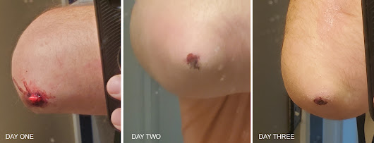
Day 1- pain levels 8/10. Moderate swelling and decrease left elbow ROM.  Unable to lift or use left upper extremity with ADLS.  PEMF treatment consisted of an undisclosed brand for 15 min at an intensity of 8.5 twice a day (one in morning one at night)

 Day 2- Pain levels 3/10, with no swelling and full ROM in the left elbow.  Able to use left upper extremity with most activities with mild pain (3/10).  Day 2 treatments same as day 1.

Day 3- pain levels 0/10 significant healing in skin injury.  Able to achieve full ROM in left elbow without pain or restrictions.  Full strength returned. Same treatment as previous days.  

Day 4 -  fully recovered to full activates without restrictions.  Mild scabbing over left elbow


EPILOGUE
During this treatment period the subject did not take any medications-prescribed or over the counter for pain.  He did not use ice or compression to the injured area.  In order to obtain clearer results his only treatment was PEMF for 4 days.  A normal recovery time of weeks was decreased to days.  The cost is minimal and full recovery was achieved.   While traditional treatment contain value, the increasing use of alterative options is gaining in popularity because of its effectiveness and safety.  Maximizing your body’s own recovery capabilities instead of medications (with possible side effects) is a far more efficient way to recovery from injuries. 

**Always consult with a trained medical professional before using any treatment.  Results may vary***


ABOUT THE CONTRIBUTORS
ROBERT L. BARD, MD, PC, DABR, FASLMS - Dr. Bard is recognized for his specialized work in advanced cancer diagnostic imaging. He co-founded the 9/11 CancerScan program to bring additional diagnostic support to all first responders from Ground Zero. His main practice in midtown, NYC (Bard Diagnostic Imaging- www.CancerScan.com) uses the latest in digital imaging technology and has been also used to help guide biopsies and in many cases, even replicate much of the same reports of a clinical invasive biopsy. Imaging solutions such as high-powered sonograms, Power Doppler Histogram, sonofluoroscopy, 3D/4D image reconstruction and the Power Doppler Histogram  are safe, noninvasive, and do not use ionizing radiation. It is used as a complement to find anomalies and help diagnose the causes of pain, swelling and infection in the body’s internal organs while allowing the diagnostician the ability to zoom and ‘travel’ deep into the body for maximum exploration. (DrRobertBard.com)


JOSH SCHUELLER, PT has dedicated his life's work to the support and treatment chronic pain and disorders with non-invasive, safe, effective treatment solutions. He is the current VP of Clinical Operations and Business Development at AxioBionics LLC. and the Clinical Director for Orthopedic Physical Therapy Clinics (Rockford, MI).  As an active member of APTA, he holds advanced certification in Physical Therapy treatment techniques including the McKenzie method of patient empowerment.  He has over 20+ years experience in the treatment of neurological conditions such as Spinal Cord Injury, Traumatic Brain Injury, CVA, Cerebral Palsy etc.  In 2021, Josh is elected a clinical advisory role and educational advocacy for IPHA (Integrated Pain Healers Alliance) and has published research articles in pain management. 


Dr. JERRY DREESSEN is both Trauma Team Qualified and Hospital Qualified treating chiropractor in Mountlake Terrace, WA. He is the Executive Director for the Association of PEMF Professionals- the largest professional PEMF organization in the United States. AOPP attracts the most principled and accomplished PEMF user- setting standards by providing certification for all PEMF users, as well as Continuing Education (CEU's) for all members to maintain best practices and techniques. (www.backtoaction.com)


Disclaimer: The information (including, but not limited to text, graphics, images and other material) contained in this article is for informational purposes only. No material on this site is intended to be a substitute for professional medical advice or scientific claims. Furthermore, any/all contributors (both medical and non-medical) featured in this article are presenting only ANECDOTAL findings pertaining to the effects and performance of the products/technologies being reviewed - and are not offering clinical data or medical recommendations in any way. Always seek the advice of your physician or other qualified health care provider with any questions you may have regarding a medical condition or treatment and before undertaking a new health care regimen, never disregard professional medical advice or delay in seeking it because of something you read on this page, article, blog or website.


Friday, February 18, 2022

Groundbreaking New Solution for Incontinence: Meet ELITONE

INTRODUCTION
Health and Healing 101 is proud to review a brilliant innovation addressing female incontinence - a most widely prevalent health and medical concern for women. As a team with extensive experience in medical device design and entrepreneurship, co-founders Gloria Kolb and Eric Kolb embarked on development of a wearable medical device called ELITONE to provide a non-intrusive incontinence treatment, capturing the attention of an expanding number of clinicians in the US.  Team Kolb shares their valuable insights on the creation of this remarkable device, while also sharing the process of achieving regulatory success for product launch.  For any aspiring developers of innovation, this interview promises to be a valuable educational tour depicting the journey of how medical technologies come to market.

Product details (sourced from https://elitone.com/)
ELITONE® is FDA cleared to treat stress urinary incontinence (SUI) and is the first transcutaneous pelvic floor muscle stimulation available in the US. The ELITONE device:
- Use externally applied electrical muscle stimulation to exercise the pelvic floor muscles
- Performs pelvic floor muscle exercises more effectively than many women can do alone
- Requires little to no instruction from a healthcare provider.
- Can be used at home, under clothing, making it easy for women to continue their everyday tasks during treatment.
- Is ideal for women with mild to moderate symptoms who would benefit from pelvic floor muscle training. 
* This makes ELITONE easy, external, and effective, which drives high levels of patient adoption and compliance. 


THE ROAD TO PRODUCING A MEDICAL DEVICE 
From an interview with CTO Eric Kolb

One in three women have incontinence. Most of them do not know that there are treatment options and they resign themselves to a life of pads and eventually diapers. Those who do explore their options quickly realize that the predominant solutions are intra-vaginally administered treatments, which many women are hesitant to pursue. 

We developed ELITONE to be externally applied, making it more comfortable and convenient for women to administer at-home. It brings recovery to women with mild-moderate stress incontinence, and in a form factor that is easy to apply.  Treatment takes only 20 minutes a day, 5x/week, for 6-12 weeks.  You can use ELITONE while you're doing other things. You can put it on and go back to work. You can get ready in the morning, sit and talk with the kids, or walk the dog. It really doesn’t interrupt your daily routine.

ORIGINS
Gloria and I started the company and made the first prototypes in our dining room. We are both engineers, both with strong backgrounds in medical device product design. Although our skill sets have a lot of overlap, I tended to work more on design, manufacturing, and quality systems, while Gloria focused on funding, clinical research, and business operations. Over time, we hired other talented people and divested some of those responsibilities to others. 

Research and development was largely funded through SBIR grants from the National Science Foundation, which started in 2015. We also received federal funding from NIH and DOD. ELITONE received FDA clearance in 2019 and we’ve been working since then to create market awareness and generate sales.

FDA
Devices that are new and higher risk are designated as Class 3 and undergo an FDA “approval” process. Moderate risk devices are designated Class 2 and follow a “clearance” process where FDA reviews them against an existing technology (i.e. a predicate). This is referred to as the 510(k) process. Some, but not all, 510(k) applications include clinical data. In the ELITONE submission we provided data that is now published in the Journal of Women’s Health Physical Therapy [link].

I've worked for big companies and small companies, and have had been through the FDA process many times. This allowed us to prepare the submissions and interact with FDA directly, without use of consultants. With ELITONE, the review was slowed because we were initially required to follow the De Novo process, a special process for relatively low-risk devices that don’t have a clear predicate. As we were nearing the end of that process another device entered the market that could be used as a predicate, so we needed to resubmit our application referencing this new predicate. It delayed us by a few months, but we are happy to have that part of development behind us. 

IRB
Muscle stimulators are designated in FDA guidance documents as non-significant risk devices that don’t require an IDE (Investigational Device Exemption) to conduct clinical research. So we use a third-party IRB firm to clear our protocols, and then conduct the clinical studies ourselves. Since ELITONE is an over-the-counter use-at-home device we take a pragmatic approach to study design. This means recruiting participants and administering treatment in ways that best match how our customers use the product. 

MARKETED AS A MEDICAL DEVICE
We have indications for both over-the-counter use and prescription use.  It's actually a lot harder to get the over-the-counter indication because you have to perform usability studies to demonstrate that potential customers can self-identify as appropriate users, that they understand the contraindications, that the Instructions for Use are adequate and correctly interpreted without a clinician's intervention, and that the patients can safely administer the treatment. 

Raising awareness of this new technology within the medical community is largely through medical conferences like the International Incontinence Society, physical therapy conferences, OBGYN conferences, etc.  These are great for having meaningful conversations with healthcare providers. Because COVID-19 limited our ability to meet with clinicians face-to-face, we've been more focused on direct-to-consumer marketing, which means a lot of internet search advertising. This approach is actually very effective because women often don't speak with their doctor about incontinence. Rather, they search online for answers to "Why am I leaking?" and "What are the solutions?"  

TYPES OF INCONTINENCE
Our current Indication for Use is for treating female stress urinary incontinence, which represents about half women with incontinence. Stress incontinence is leaking associated with coughing, sneezing, exercising, or any other increase in abdominal pressure that causes urine to leak. It is primarily affected by pelvic floor muscles, and often starts after childbirth. If untreated, it worsens as the woman ages. Thankfully, with treatment like ELITONE, many of these women can get back to living life without leaks.

Urge incontinence is associated with a sudden sensation that you need to urinate. If you’re able to make it to the bathroom without leaking it’s called Overactive Bladder. If you can’t control the urge, resulting in an accident, it’s called urge incontinence. Here, the issue is with the how nerves in the bladder are stimulated. It affects women and men as they get older, usually starting in their sixties or seventies.

THE FUTURE
We are actively working to offer new products for the other types of incontinence. We look forward to being able to offer a solution to all incontinence suffers who learn about our technology. We are actively raising capital to expand our marketing activities, and have regular conversations with potential strategic partners, including both traditional medical device companies and consumer products companies. We look forward to working with an organization to help us scale and make ELITONE widely available to the one in three women who have incontinence. 


Disclaimer: The information (including, but not limited to text, graphics, images and other material) contained in this article is for informational purposes only. No material on this site is intended to be a substitute for professional medical advice or scientific claims. Furthermore, any/all contributors (both medical and non-medical) featured in this article are presenting only ANECDOTAL findings pertaining to the effects and performance of the products/technologies being reviewed - and are not offering clinical data or medical recommendations in any way. Always seek the advice of your physician or other qualified health care provider with any questions you may have regarding a medical condition or treatment and before undertaking a new health care regimen, never disregard professional medical advice or delay in seeking it because of something you read on this page, article, blog or website.

Monday, February 7, 2022

What do you know about PEDIATRIC MYOCARDITIS?

As part of Heart Health Awareness Month, the Integrative Pain Healers Alliance, the Angiofoundation,  the editors of Prevention101 and NYCRA-news joined hands with advocacy groups like the For Elysa Foundation to form "Mommies on a Mission" to promote Pediatric Myocarditis awareness in pursuit of the national diagnostic movement.

Saving Lives Through Advocacy & Research:
FOR ELYSA FOUNDATION

Around the first week of Feb, 2022, our clinical diagnostic researcher, Dr. Robert Bard launched his PediatricScan.com 2.0 in NYC‐ which included a Pilot program for Myocarditis Screening through the use of advanced Doppler Ultrasound Imaging.  To establish the clinical network for this program is to connect with ICU specialists & Cardiologists as well as all associations supporting Myocarditis research.

We met the directors of a remarkable national advocacy foundation called FOR ELYSA FOUNDATION‐ a non‐profit organization dedicated to promoting Education, providing Light, and supporting Research in the areas of viral myocarditis and pediatric sudden cardiac arrest. (www.ForElysa.org). Mrs. Jana Rojas and husband Jaime Rojas from Kansas City developed this organization inspired by the loss of her vibrant little girl,  Elysa Louise Rojas who passed away at the tender age of two years old. " In Elysa’s case, a common childhood virus was responsible for her myocarditis. The virus either attacked Elysa’s heart directly or caused her immune system to attack her heart muscle in a “friendly fire” fashion while trying to fight the virus. The inflammation in her heart increased drastically and very quickly to the point of sudden cardiac arrest. Doctors and scientists do not fully understand the mechanisms within the body that cause a virus to “go haywire” in the immune systems of individuals with myocarditis. There is currently no way to predict when/if this will occur."

The FOR ELYSA FOUNDATION is one of our first advocacy friends in pursuit of bringing national awareness and supportive clinical research for myocarditis diagnostics and prevention. According to the ForeElysa.org website, Myocarditis is a disease marked by inflammation and damage of the heart muscle. There are many causes of myocarditis, including viral infections, autoimmune diseases, environmental toxins, and adverse reactions to medications. The most common cause of myocarditis in North America is viral infections. Myocarditis usually attacks otherwise healthy people. It is believed that 5 to 20% of all cases of sudden death in young adults are due to myocarditis. Although the exact incidence of myocarditis is not known, it is estimated that approximately 343,000 people die of myocarditis and its major complication, cardiomyopathy, each year. The prognosis is variable but chronic heart failure is the major long term complication. Myocarditis and the associated disorder of idiopathic dilated cardiomyopathy are the cause of approximately 45% of heart transplants in the United States.  

All materials in this section are published with express consent from The For Elysa Foundation.  For complete Information, visit www.FORELYSA.org


HOW & WHEN TO CHECK FOR PEDIATRIC MYOCARDITIS
By: Jana Rojas

The tricky thing with myocarditis being virally mediated is that Elysa could have had a heart scan a week before she died (the day before she contracted the virus that wrecked her heart), and it would have been normal. I am hesitant to insinuate that imaging could "clear" a patient and provide a clean bill of health without noting that this can and does occur spontaneously after viral infections, and so testing while ill or post-virally is actually the key message and window of opportunity for myocarditis detection. In my mind, the primary role for cardiac diagnostic imaging as it related to myocarditis specifically would be for: 

1) acutely ill children in ED/urgent care/hospital inpatient settings

2) children exhibiting the signs and symptoms you have outlined (fainting, sudden fatigue, shortness of breath, chest pain, palpitations), 

3) after known Covid or other viral infection with prolonged or delayed healing (ie ongoing fatigue, shortness of breath, etc)  

4) and possibly PRE-PARTICIPATION SPORTS PHYSICALS. The pre participation screenings would be enhanced cardiac screenings in general to ideally pick up congenital heart defects and other concerns as well as myocarditis. 


MedNews 101

Myocarditis Detection  Written by: Dr. Robert L. Bard
Since the advent of Covid-19 Long Haul studies in 2021, the medical diagnostic community shifted into overdrive- seeking out all available screening and examination protocols to assess health problems called POST-ACUTE SEQUELAE (PASC). One of the recent Covid-19 related headliners is the rise in cases of MYOCARDITIS in children 16 years and under. CDC Reports link the pathological impact with covid infection since it is proven that Viral infections are a common cause of myocarditis. 

Between early 2020–2021, patients with Covid-19 had nearly 16 times the risk for myocarditis[1]. According to the CDC, in a study of myocarditis cases, 2,116 (41.7%) had a history of Covid-19.  In addition, cases of myocarditis reported to the Vaccine Adverse Event Reporting System (VAERS) indicated links between Myocarditis and Pericarditis to come from the mRNA Covid-19 vaccination (especially in male adolescents and young adults) more often after the second dose.[2].

Myocarditis is defined a an inflammatory disorder of the heart muscle (myocardium) leading to cardiac dysfunction. It is also recognized as myocardial cell death [3]. Checkups for this also reviews for PERICARDITIS (the inflammation of the outer lining of the heart). Various causes of myocarditis includes: Viral Infectious including adenoviruses, echoviruses, enteroviruses like the coxsackie viruses. In addition, predisposition can occur from those with Autoimmune diseases such as Celiac disease, Churg-Strauss syndrome, Crohn disease, Kawasaki disease, lupus, rheumatoid arthritis, sarcoidosis etc. (See NIH chart for full list of causes- [4]).

STANDARDIZED DIAGNOSTICS: 
As with any critical disorder, detecting early stages of myocarditis allows for a higher opportunity to treat and even eliminate the health risk.  In children, symptoms include: Fever, Fainting, Breathing difficulties, Rapid breathing, Chest pain and Rapid or irregular heart rhythms [5]. In adults, symptoms range from chest pain, shortness of breath, at rest or during activity and fluid buildup with swelling of the legs, ankles and feet. To prevent possible heart damage, a cardiologist may order one of a umber of imaging options:

- Electrocardiogram (ECG or EKG)
- Chest X-Ray
- Heart MRI
- Blood Tests
- Doppler Ultrasound for Acute Myocarditis
- Cardiovascular MR Elastography (MRE)
- Ultrasound Elastography


See expanded details on diagnostic protocols, visit:  http://pediatricscan.com/myocarditis.html


TROUBLESHOOTING MYOCARDITIS
By Bobbi Kline, MD (Integrative Physician / Genomic Research Specialist)

As a mom, my heart grieves for parents, including Elysa’s, who suffer such devastating tragedy. It’s the worst thing you hope never happens to your child, and I truly admire parents who turn a tragedy into something positive. It requires such amazing strength, courage and grace. As a physician, I find my self immediately asking "Why do these things happen? How can we predict or prevent them?" As clinicians, we look for patterns to help guide diagnosis and treatment. We know what to expect, but sometimes they can lull us into a false sense of security. Childhood viruses, as any parent knows, are an expected part of those early years. 
But what happens when they turn out to be something more? That’s where pattern recognition is crucial. When something obviously falls outside those patterns, it’s a signal to question and go deeper. But what happens when you don’t even recognize that deviation? What if something is so uncommon or so subtle that it’s hard to detect among all the noise? Post-viral myocarditis is one of those conditions, and I’m glad to see it now in the spotlight. Raising awareness is a key first step. While COVID-19 has certainly helped to highlight this condition, it goes further than COVID. Many common childhood viruses have been implicated in causing myocarditis, but most people are completely unaware. I admit that it was not something I ever really thought about as my kids were growing up. And I am not alone. Educating clinicians as well as parents on what to look for, when to be alarmed, when to go deeper is crucial. This alone will save lives. 
But it’s only the first step. We also need better tests and tools to quickly and easily identify who is at risk, and better treatments for helping these children. This requires a multidisciplinary approach that includes better diagnostics including noninvasive technology, along with effective medications and other treatments. It also includes the burgeoning field of genomics and personalized medicine, both to provide a better understanding of the why, as well as a powerful tool to predict and prevent. For, at the heart of this, is understanding each child’s uniqueness in a way that empowers.  Two studies have been published this year that have the potential to leverage the power of DNA to identify who is at risk for developing myocarditis after a viral infection. Not only that, but also which of those children are most likely to recover, and therefore need fewer interventions, and which of those children are most at risk for sudden death and require much more intensive treatment and support. And, in today’s world, we also need the power of legislation to make sure everyone has access to this higher level of care. There is much promise to change the trajectory of this devastating illness, and it is only through advocacy such as this that it will happen. 

MedNews Extra
A Pediatric Cardiologist Talks Myocarditis & COVID-19 | Perspectives on COVID-19 Vaccine for Kids

VIDEO SPOTLIGHT 1: Dr. Matthew Elias, Pediatric Cardiologist at Children’s Hospital of Philadelphia talks Myocarditis & COVID-19 | Perspectives on COVID-19 Vaccine for Kids 

Credit: Children’s Hospital of Philadelphia

As a pediatric cardiologist at the Children’s Hospital of Philadelphia, Dr. Matt E. has treated children experiencing inflammation of the heart muscle, called myocarditis, following COVID-19 infection or receipt of COVID-19 vaccine. Dr.  Matthew Elias discusses the differences between myocarditis in these two scenarios, stating, “It's important to know that heart issues, including myocarditis, are much more common and more severe with the COVID-19 infection compared to the vaccine.” For information about COVID-19, visit http://COVIDVaccineAnswers.org

Video created by and for the Vaccine Education Center at Children’s Hospital of Philadelphia. ©2021. All rights reserved.

The Vaccine Education Center at Children’s Hospital of Philadelphia (VEC at CHOP) is composed of scientists, physicians, mothers, and fathers devoted to the study and prevention of infectious diseases. The Center was launched in October 2000 to provide accurate, comprehensive and up-to-date information about vaccines and the diseases they prevent.  For information about vaccines and vaccine safety, visit http://vaccine.chop.edu.


THE 2022 NATIONAL "ONE VOICE" FOR CHILDREN COLLECTIVE

In Feb. 2022, lead directors of The AngioFoundation, The NY Cancer Resource Alliance and publishers of PREVENTION101.org forged a collaborative outreach community project with the For Elysa Foundation (myocarditis awareness org) to form a national coalition of clinical support associations, research foundations, researchers and parent groups dedicated to the awareness of rare pediatric diseases and critical disorders. This collective concept called "MOMMIES ON A MISSION" is a unique and remarkable platform that aspires to support the many pediatric disorders that plague today's children.

Known as 'Project One", Mommies on a Mission will launch the ONE VOICE crusade to leverage new awarness response for the deadly health threat of Myocarditis in America's children. "MOMMIES ON A MISSION" is a creative title Mrs. Jana Rojas (director of For Elysa Foundation) used to describe her fellow advocates who, like her, established a dedicated benevolent program to battle pediatric myocarditis through education, awareness and to support clinical research. Even more are driven to forge standardized screening of myocarditis as part of a national prevention and early detection initiative. "Losing another child to this terrible and insidious disease is one child too many! It's time we joined hands with all groups, legislators, medical societies and anyone who can make a difference. We hope to deploy a multi-stage battle plan to address this disease and enact new solutions in every pediatrician's office!", states Dr. Noelle Cutter (Molloy College, NY Cancer Resource Alliance).


REFERENCES

1) Morbidity and Mortality Weekly Report (MMWR): Association Between Covid-19 and Myocarditis Using Hospital-based Admin Data 3/2020-1/2021) https://www.cdc.gov/mmwr/volumes/70/wr/mm7035e5.htm
3) MR Imaging of Myocardial Infarction | RSNA-Radiological Society of North America  / https://pubs.rsna.org/doi/10.1148/rg.335125722
4) The Diagnostic and Clinical Approach to Pediatric Myocarditis: A Review of the Current Literature (NCBI/NIH)  https://www.ncbi.nlm.nih.gov/pmc/articles/PMC6352488/
5) Diagnosis and Management of Myocarditis in Children (American Coll. of Cardiology) https://www.acc.org/latest-in-cardiology/ten-points-to-remember/2021/07/09/17/31/diagnosis-and-management-of-myocarditis

Thursday, February 3, 2022

PEDIATRIC MYOCARDITIS & NATIONAL DETECTION MOVEMENT (Part 1)

Written by: Dr. Robert L. Bard

Since the advent of Covid-19 Long Haul studies in 2021, the medical diagnostic community shifted into overdrive- seeking out all available screening and examination protocols to assess health problems called POST-ACUTE SEQUELAE (PASC). One of the recent Covid-19 related headliners is the rise in cases of MYOCARDITIS in children 16 years and under. CDC Reports link the pathological impact with covid infection since it is proven that Viral infections are a common cause of myocarditis. 

Between early 2020–2021, patients with Covid-19 had nearly 16 times the risk for myocarditis[1]. According to the CDC, in a study of myocarditis cases, 2,116 (41.7%) had a history of Covid-19.  In addition, cases of myocarditis reported to the Vaccine Adverse Event Reporting System (VAERS) indicated links between Myocarditis and Pericarditis to come from the mRNA Covid-19 vaccination (especially in male adolescents and young adults) more often after the second dose.[2].

Myocarditis is defined as an inflammatory disorder of the heart muscle (myocardium) leading to cardiac dysfunction. It is also recognized as myocardial cell death [3]. Checkups for this also reviews for PERICARDITIS (the inflammation of the outer lining of the heart). Various causes of myocarditis includes: Viral Infectious including adenoviruses, echoviruses, enteroviruses like the coxsackie viruses. In addition, predisposition can occur from those with Autoimmune diseases such as Celiac disease, Churg-Strauss syndrome, Crohn disease, Kawasaki disease, lupus, rheumatoid arthritis, sarcoidosis etc. (See NIH chart for full list of causes- [4])

STANDARDIZED DIAGNOSTICS
As with any critical disorder, detecting early stages of myocarditis allows for a higher opportunity to treat and even eliminate the health risk.  In children, symptoms include: Fever, Fainting, Breathing difficulties, Rapid breathing, Chest pain and Rapid or irregular heart rhythms [5]. In adults, symptoms range from chest pain, shortness of breath, at rest or during activity and fluid buildup with swelling of the legs, ankles and feet. To prevent possible heart damage, a cardiologist may order one of a number of imaging options:

Electrocardiogram (ECG or EKG)
Chest X-Ray
Heart MRI
Blood Tests
Doppler Ultrasound for Acute Myocarditis
Cardiovascular MR Elastography (MRE)
Ultrasound Elastography

See expanded details on diagnostic protocols, visit:  http://pediatricscan.com/myocarditis.html


MedNews Extra
Saving Lives Through Advocacy & Research:
FOR ELYSA FOUNDATION

Around the first week of Feb, 2022, our clinical diagnostic researcher, Dr. Robert Bard launched his PediatricScan.com 2.0 in NYC‐ which included a Pilot program for Myocarditis Screening through the use of advanced Doppler Ultrasound Imaging.  To establish the clinical network for this program is to connect with ICU specialists & Cardiologists as well as all associations supporting Myocarditis research.

We met the directors of a remarkable national advocacy foundation called FOR ELYSA FOUNDATION‐ a non‐profit organization dedicated to promoting Education, providing Light, and supporting Research in the areas of viral myocarditis and pediatric sudden cardiac arrest. (www.ForElysa.org). Mrs. Jana Rojas and husband Jaime Rojas from Kansas City developed this organization inspired by the loss of her vibrant little girl,  Elysa Louise Rojas who passed away at the tender age of two years old. " In Elysa’s case, a common childhood virus was responsible for her myocarditis. The virus either attacked Elysa’s heart directly or caused her immune system to attack her heart muscle in a “friendly fire” fashion while trying to fight the virus. The inflammation in her heart increased drastically and very quickly to the point of sudden cardiac arrest. Doctors and scientists do not fully understand the mechanisms within the body that cause a virus to “go haywire” in the immune systems of individuals with myocarditis. There is currently no way to predict when/if this will occur."


The FOR ELYSA FOUNDATION is one of our first advocacy friends in pursuit of bringing national awareness and supportive clinical research for myocarditis diagnostics and prevention. According to the ForeElysa.org website, Myocarditis is a disease marked by inflammation and damage of the heart muscle. There are many causes of myocarditis, including viral infections, autoimmune diseases, environmental toxins, and adverse reactions to medications. The most common cause of myocarditis in North America is viral infections. Myocarditis usually attacks otherwise healthy people. It is believed that 5 to 20% of all cases of sudden death in young adults are due to myocarditis. Although the exact incidence of myocarditis is not known, it is estimated that approximately 343,000 people die of myocarditis and its major complication, cardiomyopathy, each year. The prognosis is variable but chronic heart failure is the major long term complication. Myocarditis and the associated disorder of idiopathic dilated cardiomyopathy are the cause of approximately 45% of heart transplants in the United States.  

Materials in this excerpt are published with express consent from The For Elysa Foundation.  For complete Information, visit www.FORELYSA.org


HOW & WHEN TO CHECK FOR PEDIATRIC MYOCARDITIS
By: Jana Rojas
The tricky thing with myocarditis being virally mediated is that Elysa could have had a heart scan a week before she died (the day before she contracted the virus that wrecked her heart), and it would have been normal. I am hesitant to insinuate that imaging could "clear" a patient and provide a clean bill of health without noting that this can and does occur spontaneously after viral infections, and so testing while ill or post-virally is actually the key message and window of opportunity for myocarditis detection.
In my mind, the primary role for cardiac diagnostic imaging as it related to myocarditis specifically would be for: 
1) acutely ill children in ED/urgent care/hospital inpatient settings
2) children exhibiting the signs and symptoms you have outlined (fainting, sudden fatigue, shortness of breath, chest pain, palpitations), 
3) after known Covid or other viral infection with prolonged or delayed healing (ie ongoing fatigue, shortness of breath, etc)  
4) and possibly PRE-PARTICIPATION SPORTS PHYSICALS. The pre participation screenings would be enhanced cardiac screenings in general to ideally pick up congenital heart defects and other concerns as well as myocarditis. 


TROUBLESHOOTING MYOCARDITIS
By Bobbi Kline, MD (Integrative Physician / Genomic Research Specialist)

As a mom, my heart grieves for parents, including Elysa’s, who suffer such devastating tragedy. It’s the worst thing you hope never happens to your child, and I truly admire parents who turn a tragedy into something positive. It requires such amazing strength, courage and grace. As a physician, I find my self immediately asking "Why do these things happen? How can we predict or prevent them?" As clinicians, we look for patterns to help guide diagnosis and treatment. We know what to expect, but sometimes they can lull us into a false sense of security. Childhood viruses, as any parent knows, are an expected part of those early years. 

But what happens when they turn out to be something more? That’s where pattern recognition is crucial. When something obviously falls outside those patterns, it’s a signal to question and go deeper. But what happens when you don’t even recognize that deviation? What if something is so uncommon or so subtle that it’s hard to detect among all the noise? Post-viral myocarditis is one of those conditions, and I’m glad to see it now in the spotlight. Raising awareness is a key first step. While COVID-19 has certainly helped to highlight this condition, it goes further than COVID. Many common childhood viruses have been implicated in causing myocarditis, but most people are completely unaware. I admit that it was not something I ever really thought about as my kids were growing up. And I am not alone. Educating clinicians as well as parents on what to look for, when to be alarmed, when to go deeper is crucial. This alone will save lives. 

But it’s only the first step. We also need better tests and tools to quickly and easily identify who is at risk, and better treatments for helping these children. This requires a multidisciplinary approach that includes better diagnostics including noninvasive technology, along with effective medications and other treatments. It also includes the burgeoning field of genomics and personalized medicine, both to provide a better understanding of the why, as well as a powerful tool to predict and prevent. For, at the heart of this, is understanding each child’s uniqueness in a way that empowers.  Two studies have been published this year that have the potential to leverage the power of DNA to identify who is at risk for developing myocarditis after a viral infection. Not only that, but also which of those children are most likely to recover, and therefore need fewer interventions, and which of those children are most at risk for sudden death and require much more intensive treatment and support. And, in today’s world, we also need the power of legislation to make sure everyone has access to this higher level of care. There is much promise to change the trajectory of this devastating illness, and it is only through advocacy such as this that it will happen. 


1) Morbidity and Mortality Weekly Report (MMWR): Association Between Covid-19 and Myocarditis Using Hospital-based Admin Data 3/2020-1/2021) https://www.cdc.gov/mmwr/volumes/70/wr/mm7035e5.htm
3) MR Imaging of Myocardial Infarction | RSNA-Radiological Society of North America  / https://pubs.rsna.org/doi/10.1148/rg.335125722
4) The Diagnostic and Clinical Approach to Pediatric Myocarditis: A Review of the Current Literature (NCBI/NIH)  https://www.ncbi.nlm.nih.gov/pmc/articles/PMC6352488/
5) Diagnosis and Management of Myocarditis in Children (American Coll. of Cardiology) https://www.acc.org/latest-in-cardiology/ten-points-to-remember/2021/07/09/17/31/diagnosis-and-management-of-myocarditis


Disclaimer: The information (including, but not limited to text, graphics, images and other material) contained in this article is for informational purposes only. No material on this site is intended to be a substitute for professional medical advice or scientific claims. Furthermore, any/all contributors (both medical and non-medical) featured in this article are presenting only ANECDOTAL findings pertaining to the effects and performance of the products/technologies being reviewed - and are not offering clinical data or medical recommendations in any way. Always seek the advice of your physician or other qualified health care provider with any questions you may have regarding a medical condition or treatment and before undertaking a new health care regimen, never disregard professional medical advice or delay in seeking it because of something you read on this page, article, blog or website. None of the information provided should be interpreted to be or is meant to be medical advice, suggestions, or counseling.

A SURVIVOR'S PATH TO ENDO-MENTORING

By: Milcah Joy Macatiag / empowerment speaker for Endo patients I’m a Critical Care Nurse With 3 decades of experience in healthcare. My pro...